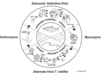
Lifecycle of Myxobolus cerebralis, KALLERT D. unpubl.
|
Lifecycle of Myxobolus cerebralis, KALLERT D. unpubl. |
Salmonid whirling disease (WD) is responsible for the great loss of rainbow trout fry (EL-MATBOULI et al.,
1992; HEDRICK et al., 1998). The high mortality makes WD an important economical issue especially in the United States.
The parasitic organism causing WD is the myxosporean Myxobolus cerebralis (HOFFMAN, 1990) (Myxozoa). The whole phylum is now
discussed to be closely related to cnidarians (USPENSKAIA & RAIKOVA, 2001; SIDDAL et al., 1995). The parasite follows a
complex life cycle with sporogony in each of the two hosts (Tubifex tubifex, Oncorhychus mykiss). M. cerebralis sporoplasms,
after leaving the fish-infective actinospore (TAM), undergo several multiplications in the epidermis and migrate along the nervous tissue to
anterior parts of the fish host, where the amoeboid sporoplasm attacks cranial cartilage. Here the trophozoids mature and produce very
durable myxospores. These have to be ingested by T. tubifex after being released from the dead fish host. Acute symptoms are
curly movements and a black tail mostly affecting young fish up to one year of age. Elder fish rarely die of WD due to proceeded
ossification.
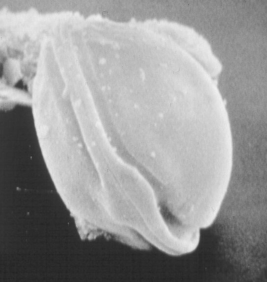
TAMs seem to be able to recognize suceptible hosts specifically (MARKIW, 1989; EL-MATBOULI et al. 1995). After contact they
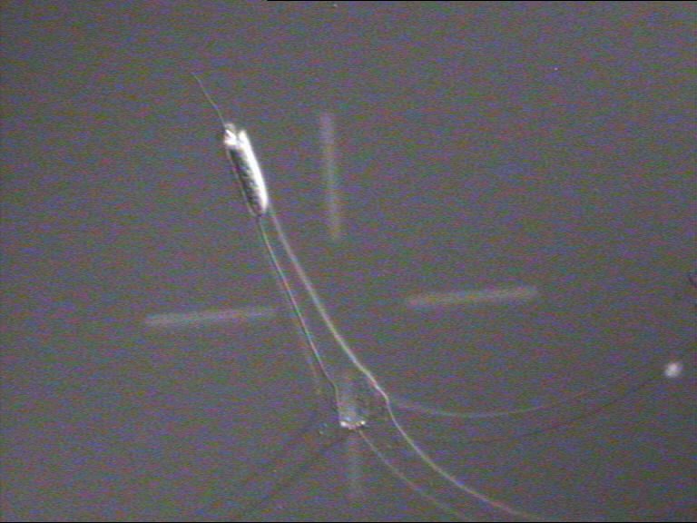
extrude polar filaments stored in 3 capsules at the end of the spore body. The physiological nature of this reaction is not understood,
and proper stimuli and triggers still remain completely unknown, though some investigational effort has been done (WAGNER, 2001).
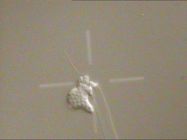
Another point of my work is the mechanism starting the invasion movements of the sporoplasm, about which also no information except
morphological work is available to date. Further knowledge about the components that cause or trigger these reactions are of great
interest for the development of suitable possibilities of field control or prophylactic therapy.
In cooperation with PD DR. DR. M. El-Matbouli (Institute for Zoology, Fishing-Biology and Fish Diseases, Ludwig
Maximilians-University, Munich) we try to isolate the components from trout mucus, that are important for host recognition by various
biochemical techniques. Several in vitro bioassays for a quantitative evaluation of these reactions of the TAMs have been developed,
to obtain scientific data.
|
|
|
||||
Wilfried Haas, Dennis Kallert, Jennifer Borrelli, Sabine Ponader
![]() Homepage
Homepage